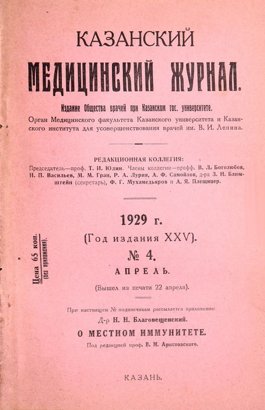
Grashey K. Atlas typischer Röntgenbilder vom normalen Menschen ed., Lehmann, Munchen, 1928, price 26 M. in hardcover
- Authors: Gasul R.Y.
- Issue: Vol 25, No 4 (1929)
- Pages: 453-454
- Section: Articles
- Submitted: 13.09.2021
- Accepted: 13.09.2021
- Published: 15.04.1929
- URL: https://kazanmedjournal.ru/kazanmedj/article/view/80016
- DOI: https://doi.org/10.17816/kazmj80016
- ID: 80016
Cite item
Full Text
Abstract
New expanded and supplemented 5th ed. the famous atlas of the German radiologist Grashey does not need any special recommendation. The normal X-ray picture is the basis for the study of pathological. Insufficiently thorough acquaintance with the normal picture was the source of sometimes very gross radiological errors. Too often the radiologist saw pathology where at least there was only a variant, and for the most part, the norm. As we accumulated our experience, as we became familiar with the variations of the depicted organs, not only depending on the constitutional characteristics of the organism and the individual, but also on the position of the given organ in relation to the tube and plate, we learned to avoid mistakes. This atlas of the normal skeleton in x-ray has character and guidelines. The first 97 pages contain: physical fundamentals (instrumentation, exposure, fixators, hoods, protective devices, photographic equipment), practical basics of centering and perspective, excellent drawings and diagrams of patient positioning with various images, diagrams of normal ossification, options and valuable instructions for analyzing radiographs. In the second section, on 140 pages of the best paper, there are 234 excellent positive radiographs that are not inferior to the originals and were made according to a special method (Glanzdruck). These impressions allow a detailed analysis of each image, the selection of which is an excellent material for comparison, equally useful for a beginner, and a specialist-radiologist, and an orthopedic surgeon.
Keywords
Full Text
Новое расширенное и дополненное 5 изд. известного атласа германского рентгенолога Grashey не нуждается в особой рекомендации. Нормальная рентгеновская картина является основой при изучении патологической. Недостаточно основательное знакомство с нормальной картиной являлось источником порою очень грубых рентгенологических ошибок. Слишком часто рентгенолог видел патологию там, где по меньшей мере имелся лишь вариант, а большей частью—норма. По мере накопления нашего опыта, по мере ознакомления с вариациями изображаемых органов не только в зависимости от конституциональных особенностей организма и индивидуума, но и от положения данного органа по отношению к трубке и пластинке,—мы научились избегать ошибок. Настоящий атлас нормального скелета в рентгеновском изображении имеет характер и руководства. Первые 97 стр. содержат: физиотехнические основы (инструментарий, экспозиция, фиксаторы, бленды, защитные приспособления, фототехника), практические основы центрировки и перспективы, прекрасные рисунки и схемы установки больного при различных снимках, схемы нормального окостенения, вариантов и ценных указаний для анализа рентгенограмм. Во втором отделе имеются на 140 стр. лучшей бумаги 234 прекрасных позитива рентгенограмм, не уступающих оригиналам и сделанных по особому методу (Glanzdruck). Эти оттиски позволяют детальный анализ каждого снимка, подбор которых представляет прекраснейший материал для сравнения, одинаково полезный и начинающему, и специалисту-рентгенологу, и ортопеду-хирургу.
About the authors
R. Ya. Gasul
Author for correspondence.
Email: info@eco-vector.com
Russian Federation
References
Supplementary files


