Neurotization of the facial nerve as an effective way to treat paralysis of mimic muscles
- Authors: Skaliitchouk B.V.1, Gaivoronsky A.I.1,2, Vinogradov V.V.1, Svistov D.V.1
-
Affiliations:
- Military Medical Academy named after S.M. Kirov
- St. Petersburg State University
- Issue: Vol 104, No 1 (2023)
- Pages: 134-143
- Section: Clinical experiences
- Submitted: 24.12.2021
- Accepted: 29.09.2022
- Published: 01.02.2023
- URL: https://kazanmedjournal.ru/kazanmedj/article/view/90970
- DOI: https://doi.org/10.17816/KMJ90970
- ID: 90970
Cite item
Abstract
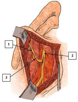
Pathologies of the facial nerve are one of the most common types of pathology of the peripheral nervous system. In the structure of lesions of the cranial nerves, this pathology occupies the first place. The clinical picture of facial nerve damage of various genesis is rather monotonous and manifests itself as persistent paresis or paralysis of the facial muscles. The literature describes a large number of different highly effective techniques aimed at restoring the function of the facial nerve and mimic muscles, examples of which are numerous conservative and surgical methods for the treatment of facial nerve neuropathy. The review presents the most common method of mimic muscles paralysis surgical treatment — facial nerve neurotization. The essence of this surgical intervention is in suturing to the affected facial nerve the trunk or a portion of individual fibers of the intact nerve-neurotic, which can be the hypoglossal, masticatory, phrenic, accessory, glossopharyngeal nerves, as well as the descending branch of the hypoglossal nerve and the anterior branches of the C2–C3 cervical nerves. Currently, options for the combined use of various donor nerves and autoextensions are gaining popularity among neurosurgeons, due to more favorable results in restoring the function of the facial nerve, as well as with an individual approach to each patient. The main stages of neurotization of the facial nerve include the isolation and intersection of the facial nerve, the isolation and intersection of the trunk or individual fibers of the neurotizer, the execution of the suture of the nerve in the “end to end” or “end to side” method. Particular attention should be paid to the most innovative method of facial nerve neurotization — facial nerve cross-plasty, during which an anastomosis between the damaged and intact facial nerves using autotransplants from the gastrocnemius nerve or a free muscle graft, including the tender muscle and the anterior branch of the obturator nerve is performed. The process of restoring facial nerve function and regressing characteristic symptoms takes a long period of time and requires specialized restorative treatment.
Full Text
Facial nerve neuropathy (FNN) is a condition that causes paresis or paralysis of the facial muscles and impairs parasympathetic innervation of the eye, resulting in significant aesthetic defects and reduced quality of life. According to reports, the incidence of FNN is estimated to be 1.3–5.4 cases per 10,000 population [1–3]. Damage and diseases of the facial nerve (FN) are the most common lesions of cranial nerves and the second most common in the peripheral nervous system [4, 5].
The etiological factors of FNN are diverse, and M. May and D.G. Klein classify them into obstetric, traumatic, neurological, infectious, metabolic, neoplastic, toxic, iatrogenic, and idiopathic causes [5].
Neurosurgeons primarily treat mechanical injuries of the facial nerve in cases of craniocerebral and craniofacial trauma and iatrogenic injuries resulting from the removal of posterior cranial fossa neoplasms (most commonly vestibular schwannomas) and parotid salivary gland tumors. They also perform surgical interventions on the peripheral part of the auditory analyzer and the middle ear [2, 5, 6]. M.H. Hofman et al. [7] reported that FNN is a postoperative complication in 10% of cases following resection of VIII cranial nerve neurinoma in a study of 1989 patients. In cases of temporal bone fractures, FNN occurs in 7–10% of cases [8].
The severity of FN dysfunction and the effectiveness of FNN treatment are commonly assessed using J.W. House and D.E. Brackmann scale, the M. May scale and electroneuromyography [6–8].
Surgical aids are effective for persistent FN dysfunction, which include interventions on the FN to restore its conduction and plastic surgeries (static and dynamic) performed on the skin, muscles, and tendons of the face. The aim of plastic surgeries is to reduce or remove cosmetic defects and replace the function of paralyzed muscles [5, 9–11].
Treatment of FN is relevant for various surgical, therapeutic, and rehabilitation specialists because of its topographical and anatomical features and the frequency of FNN occurrence during various surgical interventions [12].
The Russian [11, 13, 14] and foreign [4, 5, 15] literature has been devoted to the issue of selecting an effective surgical treatment of FNN. A.I. Nerobeev and C.M. Shurgai have developed a comprehensive surgical method for treating FNN, which consists of decompression and neurolysis of the FN, neurotization, direct neurorrhaphy, transposition of FN branches, replacement of diastasis with autografts, and cross-facial nerve autotransplantation [9].
This review presents methods for FN neurotization as a surgical treatment option for mimicking muscle paralysis. Neurotization of the FN is a surgical technique used to restore the function of denervated facial musculature, primarily to restore movement, which is achieved by sewing the central end of another nerve trunk, located near or far away and considered functionally less significant, into the peripheral end of the FN. The nerve in question is referred to as the “neurotizer nerve” in the Russian literature [16–18] and as the “donor nerve” in the foreign literature [2, 7].
The FN is the nerve on which reinnervation was first attempted to restore its function. Many donor nerves have been used for FN neurotization, including the hyoid, masseter, accessory, lingual, laryngeal, diaphragmatic nerves, the descending branch of the hyoid nerve, and the anterior branches of the C2–C3 cervical nerves [6, 9, 11, 16, 17].
Surgical attempts to suture the branch of the upper cervical sympathetic trunk (internal carotid nerve) to the peripheral end of the facial nerve have been described, they were found to be ineffective. Sympathectomy, when performed, was found to improve the surgical intervention. V.I. Grebenyuk [11] recommends this method only for cases of facial muscle paresis. The patients showed some revitalization of the function of their mimic muscles, resulting in tighter eyelid closure and a more symmetrical face. Some necrotizing nerves mentioned above are now considered to have only historical and exploratory significance in modern surgical approaches for treatment of FNN [11].
In 1949, Russian surgeons F.A. Poemny and F.M. Khitrov proposed a variant of diaphragmatic neurotization of FN. They observed a positive outcome in five out of six patients, but in one patient, neurotization was impossible because of the insufficient length of the neurotizer nerve [11]. In 1954, V.I. Grebenyuk addressed this problem by using a mobilizer of the diaphragmatic nerve. However, a side effect of this intervention is the frequent development of movements of the mimic musculature concomitant with breathing, requiring long-term corrective therapy [12].
At present, the accessory, hyoid, and masseter nerves are commonly used for FN neurotization. Previous works have described a combined approach using the accessory nerve and the descending branch of the hyoid nerve [6, 17–19].
The first attempt to innervate FN with an accessory nerve was conducted by T. Drobnik in 1879. Later, in 1895, the English surgeon Ch. Balance performed this intervention but obtained disappointing results [2, 11]. In Russia, V.M. Mintz performed this intervention first in 1903, followed by similar surgeries by V.N. Zerenin (1905), I.K. Spizharny (1906), and others [11]. Different methods have been described for using a separate portion of the motor fibers of the accessory nerve to the m. sternocleidomastoideus and suturing its common trunk to the FN [11]. To access this area, the anterior edge of the upper third of the nodus muscle is approached, followed by the area behind the parotid salivary gland in the region of the stylosceles foramen. The FN trunk is then isolated and crossed, after which the isolated trunks are sutured end-to-end (Fig. 1).
Fig. 1. Scheme of access to the facial and accessory nerves: 1, facial nerve; 2, accessory nerve; 3, parotid salivary gland; 4, internal jugular vein; 5, sternocleidomastoid muscle.
However, using this method always leads to consequences related to atrophic changes in the trapezius and sternoclavicular–parietal muscles. Recovery of the FN function after such surgical intervention will be demonstrated by common movements of the facial musculature with arm movements, which requires long-term retraining and rehabilitation [11, 12].
When the descending branch of the hyoid nerve is used as a neurotizer nerve, access is performed in the lateral triangle of the neck, extended toward the neurovascular bundle of the neck. The branch is then mobilized and crossed at the bifurcation level of the common carotid artery. Importantly, the diameter of the descending branch of the hyoid nerve is often significantly smaller, at only 25% of the diameter of the entire nerve [20], compared with that of the FN trunk.
For more effective neurotization, attempts are sometimes made to exclude functionally insignificant bundles from the neurotized (facial) nerve. This directs more neurotizing nerve fibers to the most functionally significant facial muscles. F.S. Govenko [16] observed that this technique obtains the best results in cases of FN neurotization by the descending branch of the hyoid nerve. In such cases, the temporal, zygomatic, and cheek branches, along with the marginal branch of the mandible, are reinnervated. The bundles of the FN that innervate the subcutaneous neck muscle, hyoid, and bicuspid muscles are excluded from the area of neurorrhaphy [16].
The method of FN neurotization using the anterior branches of the II–III cervical nerves includes isolating the distal part of the FN and accessing the donor nerves along the anterior edge of the upper third of the m. sternocleidomastoideus [6, 14, 17].
The abovementioned methods were presented in a series of studies by Y.V. Tsymbaliuk et al. [6], which included 172 patients. At the time of surgical intervention, FN dysfunction reached the VI degree according to the House–Brackman scale, corresponding to total paralysis of the mimic muscles. The results of FN neurotization showed that 85.4% of cases (147 patients) achieved a satisfactory result corresponding to a degree of II–III according to the House–Brackman scale. In variants of FN neurotization by additive and combined methods (using several neurotizers), the success rates were 89.7% and 96.8%, respectively. However, using anterior motor branches of the II–III cervical nerves did not result in any recovery of the FN function.
However, V.V. Kaverina [14] presented two clinical observations of FN neurotization by the ventral branch of the II cervical nerve with a favorable outcome. In both cases, the patients had total unilateral paresis of the mimic muscles before surgical intervention. After neurotization, there was a gradual recovery of FN function, as verified by positive dynamics using electroneuromyography, reflected in a significant improvement of FN conduction and excitability. Based on their work, this technique is technically simple and does not result in any significant neurological deficits in the donor nerve [14].
Surgical interventions involving variants of FN neurotization by hyoid and masseter nerves are often described in the foreign literature [3, 18–20].
The technique of neurotizing the FN with the hyoid bone was first introduced by the surgeon Korte in 1903. Since then, the method has been modernized several times to preserve tongue function [21]. In 1991, M. May et al. proposed the “XII–VII jump-cable graft procedure,” which involves partially crossing the hyoid nerve and creating an end-to-side anastomosis with individual branches or the common trunk of the FN. This method is characterized by the use of one or more autografts, such as the great auricular, thoracic, and calf nerves. Provided that FN neurotization was performed within 12 months after the injury, after surgical treatment, 78% of cases showed positive results in the restoration of FN function, corresponding to the I–II degree of the May scale [21, 22].
In 1994, M.D. Cusimano et al. described the “longitudinal hypoglossal nerve technique,” which involves partially crossing the hyoid nerve fibers and longitudinally retrograde splitting fibers to further neurotize the FN (Fig. 2) [22].
Fig. 2. Method of longitudinal retrograde splitting of hyoid nerve fibers with subsequent neurotization of the facial nerve from M.D. Cusimano et al.: 1, facial nerve; 2, hyoid nerve; 3, end-to-end suture of the nerve
The next step in the development of FN neurotization options involved crossing the mastoid or tympanic segment of the FN and suturing it in an end-to-side technique to the hyoid nerve. Proposed by V. Darrouset in 1999, this technique enables anastomosis between the VII and XII nerves without tension, resulting in better preservation of tongue muscle function [23].
The use of n. hypoglossus as a nerve donor presents a major issue due to the development of tongue hemiatrophy in 100% of cases, resulting in speech and swallowing disorders [24]. To address this, the most effective location for crossing the trunk of the hyoid nerve is after its descending branch, as this preserves the innervation of the hyoid bone muscles. Surgeons used a modified surgical intervention, i.e, neurotization of the FN by the hyoid bone followed by one-stage reinnervation of the XII cranial nerve with its descending branch [24, 25].
According to M. Samii [24], no significant difference was found in the restoration of FN function when it was neurotized by the trunk of the hyoid nerve with or without reinnervation of the latter by the descending branch. However, using the first method reduces the risk of complications such as speech disorder (from 33% to 0%), tongue hemiatrophy (from 100% to 5.8%), and swallowing disorders (from 55 to 11.7%) [12, 25]. A.S. Nechaeva et al. [21] use this method and achieved positive results in 21 of 26 patients (80.7%), which corresponds to grade III–IV of the May scale. Only 7 patients (26%) showed signs of hemiatrophy of the tongue muscles, which manifested as dysarthria, but this did not affect their quality of life [23].
In a study by T.A. Dziedzic et al. [26], 84% of the 56 patients with grade VI FN dysfunction on the House–Brackman scale showed improvement to grade III. The study also evaluated the function of the hyoid nerve using the Martin scale. Ninety-three percent (53 patients) exhibited I–II degree function of the XII nerve according to the Martin scale, indicating the absence or minimal atrophy of the tongue muscles without tongue deviation. Only four patients had insignificant hemiatrophy of the tongue muscles and deviation of less than 30° (Martin III).
Several authors have emphasized the significance of timely neurotization of the FN hyoid. Surgical intervention performed within 1 to 12 months has a significant impact on the quality of reinnervation of the mimic musculature [19, 24–26]. According to Z. Gao et al., the effectiveness of reinnervation is significantly influenced by the timing of the surgery, and it should be performed no later than 12 months after nerve injury [27].
Trigeminal neurotization is a commonly used method for the neurotizing agents of FN, in which the masseter nerve is one of the most frequently employed. First described by M. Spira in 1978, this technique is performed by suturing the trunk of the masseteric nerve to the peripheral part of the FN (Fig. 3). Positive dynamics were observed in three patients who underwent this surgical intervention, with restoration of FN function and reduction of the aesthetic defect [29].
Fig. 3. Method of neurotization of the facial nerve by the masseter from M. Spira: 1, facial nerve; 2, masseter nerve; 3, end-to-end suture of the nerve; 4, parotid salivary gland
Trigeminal neurotization provides several advantages because of the topographic and anatomical relationship between the masseter nerve and the FN. The masseter nerve in the subzygomatic triangle has a comparable diameter to the FN, making it the best option for comparing the trunk or extracranial branches of the FN [30]. K. Boahenne [31] stated that the primary benefit of trigeminal neurotization is the surgical accessibility of the V and VII cranial nerves. Neurorrhaphy is conducted at the natural intersection point of the masseteric nerve and FN during surgical intervention. However, the varying depth of location in the soft tissues can result in slight tension after neurotization.
A morphology study by H.D. Fournier [32] demonstrated that the number of motor neuron axons in the masseter nerve is twice that of the FN, which is a favorable prognostic factor for the speed and quality of FN function recovery, as well as the high self-regenerative activity and myoneurotization of the intrinsic masseter muscle in case of damage to its epineurium [33].
Several studies by Russian and foreign researchers suggested a common embryonic origin of the V and VII cranial nerves. This proximity of the nuclei of these nerves results in central connections between the nucleus mesencephalicus n. trigeminus and nucleus n. facialis, as well as trigeminal-facial peripheral connections between the branches of the two regional nerves [9, 14, 30, 32, 33]. Clinically, these data are characterized by a reduced risk of developing concurrent movements and a more complete formation of nerve connections. The function of FN is regulated by the middle cerebral nucleus of the trigeminal nerve [12]. Thehe literature describes cases where mimic muscle activity has recovered independently after extracranial trauma to the FN without any surgical or therapeutic intervention. Facial muscle movements are associated with masticatory movements [34].
In work by A.V. Zotov et al. [12], the efficacy of trigeminal neurotization was evaluated in 17 patients six months after the surgical intervention and noted that 16 out of 17 patients (94.1%) showed recovery of FN function to the III–IV degree according to the House–Brackman scale, whereas one patient (5.9%) did not show any recovery. The study found that patients who underwent neurotization within 2 months of paralysis had the best results. In a series of studies conducted by F. Bigliolli et al. [35], 31 of 34 patients (91.2%) showed positive results from trigeminal neurotization. Successful reinnervation was observed on average 6 months after surgery, as evidenced by the restoration of facial symmetry.
Hontanilla et al. [36] compared the effectiveness of FN neurotization with that of hyoid and masseter nerves and observed that these methods are comparable because the restoration of FN function occurred in most cases. However, when the masseter nerve was used as a neurotizing agent, reinnervation of the FN occurred earlier with a better aesthetic effect.
Cross-face nerve grafting (facial-facial anastomosis) is currently one of the most innovative surgical treatments for FNN. Proposed by L. Scaramella in 1971, this technique involves selectively crossing some branches of the intact FN and suturing them to the branches of the affected FN. However, its use is restricted because of the limited length of branches and restricted movements of the mimic muscles after neurotization [37].
In 1976, K. Harii et al. proposed the technique of using a free muscle graft, including the m. gracilis and r. anterior n. obturatorii, to repair FN damage. The occipital nerve was sutured end-to-end to the masseter nerve on the affected side. Then, using an autograft from the calf nerve, the nerve was sutured end-to-end with the cheek branch of the intact FN, including the locking nerve in the free muscle graft [38, 39].
The most recent modification of FN cross-plasty is neurorrhaphy, which was proposed in 1992 by F. Vitebro et al. [40]. This method involves end-to-side neurorrhaphy without violating the integrity of the donor nerve. During the surgical intervention, the autograft n. suralis is used with the cheek and zygomatic branches of the FN on both sides of the face without crossing them. In modern practice, three autografts are most commonly used for the muscles of the eye, cheek, and mouth circumference. The advantage of this method is the ability to use multiple autografts to neurotize several branches simultaneously, which may reduce the likelihood of premature muscle atrophy. However, currently, cross-plasty of the FN is seldom used.
This method is characterized by the homogeneity of the function of the donor and recipient nerves, which can result in little to no difference in muscle contraction after surgery. However, axon sprouting in the autograft can take a long time, potentially leading to irreversible degeneration of the mimic muscles.
Modern literature describes various methods of combining neurotization and cross-plasty of the FN and performing cross-plasty in multiple stages [41, 42].
J.M. Kim et al. [41] observed 49 patients who underwent surgical treatment using two different techniques.
Group 1, consisting of 18 patients, underwent a two-stage surgical intervention, which involved cross-plasty of the cheek branch of FN and transplantation of a free flap of the m. gracilis. During the first stage, an autograft from the calf nerve was used to form an anastomosis between the intact and damaged cheek branches to repair the damaged FN branch. The second stage involved the implantation of a free vascular-muscular flap from a thin muscle, which was performed on average 10 months after the first stage.
Meanwhile, group 2, consisting of 31 patients, underwent a one-stage surgical intervention using the “double-innervated free gracilis muscle transfer” technique. This technique involved the formation of an anastomosis between the branch of the muscle flap’s locking nerve and the masseteric nerve in an end-to-end fashion, followed by neurorrhaphy between the r. buccalis of the intact FN and the nerve of the muscle flap using an autograft from the calf nerve.
Based on the results of the first method, a restoration of symmetry in the muscles of the mouth circumference at rest was observed. Meanwhile, the second method preserved the symmetry in the same muscle group both at rest and when smiling. However, surgical interventions of this nature have significant drawbacks, such as the possibility of restoring only a single FN branch’s function and the complexity of the surgical technique. Thus, cross-facial plasty methods show promise for treating FNN but require further development.
Conclusions
Currently, FN neurotization is a frequently used surgical intervention for treatment of paralysis and paresis of the facial muscles. The literature describes different FN neurotization techniques, including the use of one or more neurotizing nerves, autografts, cross-facial plasty techniques, and free muscle flaps. The range of surgical aids available for treatment of FNN allows the surgeon to select the most suitable neurotization option for each patient, thus improving the effectiveness of FN function restoration.
Furthermore, all known variants of FN neurotization have several disadvantages, including the inability to achieve full efficiency and the development of a certain degree of neurological deficit caused by the crossing of the intact neurotizing nerve. Few works on comparative analysis of the effectiveness of different methods of FN neurotization are found in the literature. Thus, performing an anatomical and clinical analysis to compare the primary techniques of FN neurotization is recommended.
Authors’ contribution. A.I.G. and D.V.S., concept and design of the study, editing; B.V.S., concept and design of the study, writing the text; V.V.V., collection and processing of material, writing the text.
Funding source. The study received no sponsorship support.
Conflict of interest. The authors declare no conflict of interest for the submitted article.
About the authors
Bogdan V. Skaliitchouk
Military Medical Academy named after S.M. Kirov
Author for correspondence.
Email: bogdan_skaliitchouk@mail.ru
ORCID iD: 0000-0002-6024-8142
student
Russian Federation, St. Petersburg, RussiaAlexey I. Gaivoronsky
Military Medical Academy named after S.M. Kirov; St. Petersburg State University
Email: don-gaivoronsky@ya.ru
ORCID iD: 0000-0003-1886-5486
M.D., D. Sci. (Med.), Prof., Depart. of Neurosurgery
Russian Federation, St. Petersburg, Russia; St. Petersburg, RussiaVyacheslav V. Vinogradov
Military Medical Academy named after S.M. Kirov
Email: ulytreack@gmail.com
ORCID iD: 0000-0001-5930-3805
student
Russian Federation, St. Petersburg, RussiaDmitriy V. Svistov
Military Medical Academy named after S.M. Kirov
Email: dvsvistov@mail.ru
ORCID iD: 0000-0002-3922-9887
M.D., Cand. Sci. (Med.), Assoc. Prof., Head of Depart., Depart. of Neurosurgery
Russian Federation, St. Petersburg, RussiaReferences
- Sektsiya StAR “Assotsiaciya chelyustno-litsevykh khirurgov i khirurgov-stomatologov”. Klinicheskiy protokol meditsinskoy pomoshchi patsientam s neyropatiey litsevogo nerva. (Section STAR “Association of Maxillofacial Surgeons and Dental Surgeons”. Clinical protocol of medical care for patients with neuropathy of the facial nerve.) M.; 2014. 39 р. (In Russ.)
- Reich SG. Bell's Palsy. Continuum (Minneap Minn). 2017;23(2, Selected Topics in Outpatient Neurology):447–466. doi: 10.1212/CON.0000000000000447.
- Danny J, Revenaugh PC. Facial reanimation: an update on nerve transfers in facial paralysis. Curr Opin Otolaryngol Head Neck Surg. 2019;27:231–236. doi: 10.1097/MOO.0000000000000543.
- Spencer CR, Irving RM. Causes and management of facial nerve palsy. Br J Hosp Med (Lond). 2016;77(12):686–691. doi: 10.12968/hmed.2016.77.12.686.
- Chang YS, Choi JE, Kim SW, Baek SY, Cho YS. Prevalence and associated factors of facial palsy and lifestyle characteristics: Data from the Korean National Health and Nutrition Examination Survey 2010–2012. BMJ Open. 2016;6(11):e012628. doi: 10.1136/bmjopen-2016-012628.
- Tsymbaliuk IaV, Tsymbaliuk VI, Tretyak IB, Medvediev VV, Gurianov VG, Gatskiy AA, Petriv TI. Comparative analysis of various types of neurotization as a method of surgical treatment of peripheral facial paresis. Novosti Khirurgii. 2020;28(3):299–308. (In Russ.) doi: 10.18484/2305-0047.2020.3.299.
- Hohman MH, Hadlock TA. Etiology, diagnosis, and management of facial palsy: 2000 patients at a facial nerve center. Laryngoscope. 2014;124:E283–E293. doi: 10.1002/lary.24542.
- Gordin E, Lee TS, Ducic Y, Arnaoutakis D. Facial nerve trauma: evaluation and considerations in management. Craniomaxillofac Trauma Reconstr. 2015;8(1):1–13. doi: 10.1055/s-0034-137252.
- Shurgaya CM, Nerobeev AI, Karayan AC, Myaskovskaya EM. Possibilities of neuroneurotization in the treatment of patients with facial paralysis. Stomatologiya. 1995;74(4):33–39. (In Russ.)
- Kalakutskiy NV, Pakhomova NV, Petropavlovskaya OYu. Algoritm of the choice of various options of surgical techniques of treatment of patients with tumors parotid salivary gland and paralysis of mimic muscles. Mezhdunarodnyy zhurnal prikladnykh i fundamentalnykh issledovaniy. 2016;(12-9):1637–1640. (In Russ.)
- Grebenyuk VI, Chuprina YuV. Khirurgicheskoe lechenie paralichey litsevykh myshts. (Surgical treatment of paralysis of the facial muscles.) L.: Meditsina; 1964. 155 р. (In Russ.)
- Zotov AV, Rzayev DA, Dmitriev AB, Chernov SV, Moysak GI. Evaluation of short-term surgical outcomes in facial paralysis patients treated by trigeminal neurotization. Voprosy neyrohirurgii. 2016;(4):31–39. (In Russ.) doi: 10.17116/neiro201680431-39.
- Zlotnik EI, Sklyut IA, Smeyanovich AF, Korotkeviya EA. Litsevoy nerv v khirurgii nevrinom sluhovogo nerva. (The facial nerve in surgery for acoustic neuromas.) Minsk: Belarus; 1978. 1543 р. (In Russ.)
- Kaverina VV. Regeneratsiya nervov pri neyroplasticheskikh operatsiyakh. (Nerve regeneration in neuroplastic surgery.) L.: Meditsina; 1975. 215 р. (In Russ.)
- Brown S, Isaacson B, Kutz W, Barnett S, Rozen SM. Facial nerve trauma: Clinical evaluation and management strategies. Plast Reconstruct Surg. 2019;143(5):1498–1512. doi: 10.1097/PRS.0000000000005572.
- Goven'ko FS. Khirurgiya povrezhdeniya perifericheskikh nervov. (Surgery for damage to peripheral nerves.) Saint-Petersburg: Feniks; 2010. 384 p. (In Russ.)
- Tsymbaliuk IV, Tretyak IB, Gatskiy OO, Luzan BM, Petriv TI, Tsymbaliuk VI. Differentiated methods for surgical treatment of patients with facial nerve injury consequences. Travma. 2019;20(6):45–52 (In Ukr.) doi: 10.22141/1608-1706.6.20.2019.186034.
- Matos Cruz AJ. Facial nerve repair. Treasure Island (FL): StatPearls Publishing; 2021. https://www.ncbi.nlm.nih.gov/books/NBK560623/ (access date: 14.11.2021).
- Sánchez-Ocando M, Gavilán J, Penarrocha J, González-Otero T, Moraleda S, Roda JM, Lassaletta L. Facial nerve repair: the impact of technical variations on the final outcome. Eur Arch Otorhinolaryngol. 2019; 276:3301–3308. doi: 10.1007/s00405-019-05638-8.
- Tarek A. Amer, Mohamed S. El Kholy. The split hypoglossal nerve versus the cross-face nerve graft to supply the free functional muscle transfer for facial reanimation: A comparative study. J Plast Reconstr Aesthet Surg. 2018;71:750–757. doi: 10.1016/j.bjps.2018.01.008.
- Nechaeva AS, Ulitin AYu, Pustovoy SV, Tastanbekov MM. Hypoglossal-facial nerve anastomosis for management of facial palsy. Russian neurosurgical journal named after professor AL Polenov. 2019;21(3):32–37. (In Russ.)
- Socolovsky M, Roberto S, Masi G. Treatment of complete facial palsy in adults: comparative study between direct hemihypoglossal-facial neurorrhaphy, hemihipoglossal-facial neurorrhaphy with grafts, and masseter to facial nerve transfer. Acta Neurochir. 2016;158;945–957. doi: 10.1007/s00701–016–2767–7.
- Yetiser S. Immediate hypoglossal-facial anastomosis in patients with facial interruption. J Craniofac Surg. 2018;29(3):648–650. doi: 10.1097/SCS.0000000000004150.
- Samii M, Alimohamadi M, Khouzani RK, Rashid MR, Gerganov V. Comparison of direct side-to-end and end-to-end hypoglossal-facial anastomosis for facial nerve repair. World Neurosurg. 2015;84(2):368–375. doi: 10.1016/j.wneu.2015.03.029.
- Venail F, Sabatier P, Mondain M, Segniarbieux F, Leipp C, Uziel A. Outcomes and complications of direct end-to-side facial-hypoglossal nerve anastomosis according to the modified May technique. J Neurosurg. 2009;110(4):786–791. doi: 10.3171/2008.9.JNS08769.
- Dziedzic TA, Kunert P, Marchel A. Hemihypoglossal-facial nerve anastomosis for facial nerve reanimation: Case series and technical note. World Neurosurg. 2018;118:460–467. doi: 10.1016/j.wneu.2018.06.217.
- Gao Z. Neurorrhaphy for facial reanimation with interpositional graft: Outcome in 23 patients and the impact of timing on the outcome. World Neurosurgy. 2019;19:1–16 doi: 10.1016/j.wneu.2019.02.124.
- Biglioli F, Frigerio A, Colombo V, Colletti G, Rabbiosi D, Mortini P, Dalla Toffola E, Lozza A, Brusati R. Masseteric-facial nerve anastomosis for early facial reanimation. Craniomaxillofac Surg. 2012;40(2):149–155. doi: 10.1016/j.jcms.2011.03.005.
- Hontanilla B, Marre D, Cabello A. Masseteric nerve for reanimation of the smile in short-term facial paralysis. Br J Oral Maxillofac Surgery. 2014;52(2);118–123. doi: 10.1016/j.bjoms.2013.09.017.
- Collar RM, Byrne PJ, Boahene KD. The subzygomatic triangle: Rapid, minimally invasive identification of the masseteric nerve for facial reanimation. Plast Reconstr Surg. 2013;132(1):183–188. doi: 10.1097/prs.0b013e318290f6dc.
- Boahene K. Reanimating the paralyzed face. F1000 Prime Rep. 2013;5–49. doi: 10.12703/P5-49.
- Fournier HD, Denis F, Papon X, Hentati N, Mercier P. An anatomical study of the motor distribution of the mandibular nerve for a masseteric-facial anastomosis to restore facial function. Surg Radiol Anat. 1997;19:241–244. doi: 10.1007/BF01627866.
- Gayvoronsky IV, Rodionov AA, Gayvoronskaya MG, Nichiporuk GI, Shashkov VA. Role of chewing muscles and temporomandibular joint in the realization of mandibula buttress function. Bulletin of the Russian Military medical academy. 2017;60(4):158–163. (In Russ.)
- Albathi M, Oyer S, Ishii LE, Byrne P, Ishii M, Boahene KO. Early nerve grafting for facial paralysis after cerebellopontine angle tumor resection with preserved facial nerve continuity. JAMA Facial Plast Surg. 2016;18(1);54–60. doi: 10.1001/jamafacial.2015.1558.
- Biglioli F, Colombo V, Rabbiosi D, Tarabbia F, Giovanditto F, Lozza A, Cupello S, Mortini P. Masseteric-facial nerve neurorrhaphy: results of a case series. J Neurosurg. 2017;126(1):312–318. doi: 10.3171/2015.12.jns14601.
- Hontanilla B, Marré D. Comparison of hemihypoglossal nerve versus masseteric nerve transpositions in the rehabilitation of short-term facial paralysis using the Facial Clima evaluating system. Plast Reconstr Surg. 2012;130(5):662–672. doi: 10.1097/PRS.0b013e318267d5e8.
- Zhang S, Hembd A, Ching CW, Tolley P, Rozen SM. Early masseter to facial nerve transfer may improve smile excursion in facial paralysis. Plast Reconstr Surg Glob Open. 2018;15;6(11):e2023. doi: 10.1097/GOX.0000000000002023.
- Vila PM, Kallogjeri D, Yaeger LH, Chi JJ. Powering the gracilis for facial reanimation: A systematic review and meta-analysis of outcomes based on donor nerve. JAMA Otolaryngol Head Neck Surg. 2020;146(5):429–436. doi: 10.1001/jamaoto.2020.0065.
- Boahene KO, Owusu J, Ishii L, Ishii M, Desai S, Kim I, Kim L, Byrne P. The multivecto gracilis free functional muscle flap for facial reanimation. JAMA Facial Plast Surgery. 2018;20(4);300–306. doi: 10.1001/jamafacial.2018.0048.
- Viterbo F. Secondary procedures in facial reanimation. In: Grotting JС, editor. Reoperative aesthetic & reconstructive plastic surgery. 2nd ed. St. Louis, Missouri: Qualty Medical Publishing; 2007. р. 859–879.
- Kim MJ, Kim HB, Jeong WS, Choi JW, Kim YK, Oh TS. Comparative study of 2 different innervation techniques in facial reanimation cross-face nerve graft-innervated versus double-innervated free gracilis muscle transfer. Ann Plast Surg. 2020;84(2):188–195. doi: 10.1097/sap.0000000000002034.
- Park Seong Oh, Ha Jeong Hyun, Kim Il‐Kug, Jin Ung Sik, Chang Hak. Single‐stage cross‐facial nerve grafting has a result equivalent to that of two‐stage cross‐facial nerve grafting. Microsurgery. 2020;40(2):175–182. doi: 10.1002/micr.30448.
Supplementary files


